
A small minority of manufacturers have experimented with uncemented TKRs. PMMA cement is also used in most total hip replacements, although there is debate about the merits of cemented and uncemented hips replacements. The components are secured using polymethylmethacrylate (PMMA) cement. Oxinium is a more modern - and more expensive - material that may be associated with a longer lasting knee replacement, although this is controversial. Other materials have been tried, including ceramic joints, but they have yet to work their way into mainstream surgery. Most operations replace both sides of the joint and use a plastic component over the tibia - referred to as ‘metal on plastic’. In my unit the average length of stay after surgery is around three to four days, although of course some patients stay on the ward for much longer than this. The operation takes between 60 and 90 minutes and is performed through a longitudinal incision down the front of the knee.


I always warn patients with valgus deformity of this at the time of consent but I have never actually had a patient develop foot drop in these circumstances. However, a valgus deformity is associated with risk of injury to the common peroneal nerve and subsequent foot drop. In some respects, the exact nature of the deformity doesn’t really matter and can usually be overcome. This can usually be improved during the knee replacement operation. For example, if a man in his 40s presents with surprisingly well-developed OA there is often clear evidence of an earlier sports injury, and possibly the scars of open meniscal surgery.Ī knee that doesn’t fully extend straight is said to have a fixed flexion deformity. When a valgus deformity accompanies an osteoarthritic picture there is often a history of injury in earlier life. The knee is vulnerable to fractures and meniscal tears sustained in sports and industrial injury. But in practice this is the classical appearance of rheumatoid arthritis. It is possible for the lateral side of the joint to erode first, giving a valgus deformity to the joint. On plain X-ray a narrowing of the joint space in the medial compartment can be seen, and when the patient holds their leg out straight it is usually possible to see a varus deformity. The articular cartilage that lines the surface of the joint gradually narrows and ultimately wears down to the underlying subchondral bone.

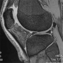
The medial jointīetween 60% and 80% of body weight is carried through the medial part of the joint and this is the most common place for the joint surface to be eroded. There are three compartments in the knee the medial, the lateral and the patellofemoral joint. Some surgeons also request ‘skyline views’ or ‘patellofemoral views’ because these may be useful in defining the state of the patellofemoral joint. When requesting plain X-rays for patients with knee pain it is useful to request a standing, weight bearing X-ray because the appearance of the knee joint can change dramatically when a patient stands up. The most common cause of pain is degenerative change in the knee, leading to osteoarthritis (OA). The patients who stand to gain the most from TKR are those with significant pain but good preoperative range of movement. Some patients also experience knee stiffness but, while TKR can alleviate this, it is harder to improve range of movement with a knee replacement than it is to reduce pain. The most common indication for a total knee replacement (TKR) is pain. Knee replacement is routine and successful, and the procedure continues to evolve. In 2015 160,000 hip and knee replacements were performed in England and Wales, with similar numbers of patients in each group. 1 CPD Credit Click here to take a test on this article and claim your certificate on MIMS Learning


 0 kommentar(er)
